baby chest x ray technique
This prediction model was transformed into a score chart. Pediatric Chest Screen 70-80 DIGITAL OPTIMUM kVp Universal CR Technique Chart using a standard 21 LgM Part View kV mAs kV mAs kV mAs Abdomen AP Grid 85 10 -15 85 20 - 25 85 30 - 40 Ankle AP 70 18 70 2 70 25 Ankle Obl 70 16 70 18 70 22 Ankle Lat 70 15 70 16 70 2 Chest -Adult AP 400 - tt -72 85 2 - 25 85 32 - 4 90 5 - 64.

Neonate Chest Supine View Radiology Reference Article Radiopaedia Org
It can help your healthcare provider see how well your lungs and heart are working.

. In this study the exposure technique of 65 kVp and 16 mAs was chosen as a reference image due to this technique being near the suitable exposure uses in pediatric chest supine AP in the DR and. If the pediatric patient can only manage. A chest X-ray is an imaging test that uses X-rays to look at the structures and organs in your chest.
A normal chest x-ray could be predicted by increasing age increasing birth weight presence of rhinitis absence of retractions and increasing arterial oxygen saturation. The aim of this study was to develop and validate a prediction. In conclusion a normal chest x-ray can accurately be.
Inspiration Penetration Rotation is part of the Lecturio course Radiology WATCH the complete course on httplect. The chest radiograph is the most common radiographic procedure performed in the imaging department and is the initial imaging modality in a patient presenting with thoracic symptoms. As radiation protection is necessary for pediatric patients it is essential to image the chest properly and avoid unnecessary repeats.
Interestingly this improved and dropped to 8 in the second month possibly as radiographers became more competent with the. This view is preferred in infant and neonate imaging whilst AP erect and PA erect views are ideal for children able to cooperate in sitting or standing 1. This video Chest X-Ray Techniques.
Certain diseases can cause changes in the structure of the heart or lungs. The ROC-area was 080 in the derivation and validation sets. Chest radiographs systematic review of chest radiographs is necessary for accurate evaluation.
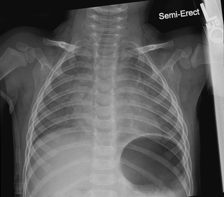
The image helps your doctor. Chest X-ray Chest X-rays produce images of your heart lungs blood vessels airways and the bones of your chest and spine. The supine chest view of the neonatal patient is a common radiographic examination when examining preterm patients 1Although not overall technically demanding the radiographer should allocate time to ensure little to no repeats are required.
Baby Chest X Ray Technique. Certain heart problems can cause changes in your lungs. Baby chest x ray technique.
Make use of digital radiography dr and needle phosphor computerised. Indications this view is preferred in infant and neonate imaging whilst ap erect and pa erect. Research surrounding the technical evaluation and technical parameters of the neonate chest x-ray is limited with a.

Pediatric Chest Supine View Radiology Reference Article Radiopaedia Org

Pedia Poser For Xray Imaging
Underinspiration And Poor Positioning Mimicking Lung Pathology In A Pediatric Patient Radiology Case Radiopaedia Org
Effect Of Changing X Ray Tube Voltage Kv Radiology Suny Upstate Medical University

Pediatric Chest X Ray In Covid 19 Infection European Journal Of Radiology
2
X Rays And Unshielded Infants Raise Alarms The New York Times
2

Chest Undergraduate Diagnostic Imaging Fundamentals

Diagnostic Imaging
2

Diagnosis Of Other Lung Conditions In Premature Babies

Pediatric Chest Horizontal Beam Lateral View Radiology Reference Article Radiopaedia Org
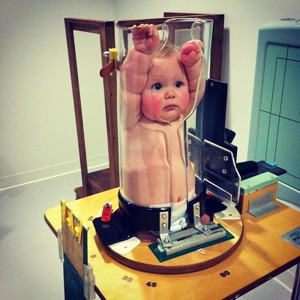
Ce4rt Guide For X Ray Techs To Immobilize Pediatrict Patients

Diagnosis Of Other Lung Conditions In Premature Babies
Effect Of Changing X Ray Tube Voltage Kv Radiology Suny Upstate Medical University

Pedia Poser For Xray Imaging

Approach To Pediatric Chest X Rays Youtube

Diagnosis Of Other Lung Conditions In Premature Babies